Applications of Volume Rendering
This page provides links to publications describing some of my collaborative activities involving the application of volume rendering methods to visualization problems in varied areas, including: cardiac lead implantation, combustion simulation, confocal microscopy, and radiation therapy treatment planning.
|
Unleaded: The Fluoroless 3D Lead Implant, Toby Markowitz, Phillip Falkner, Chad Giese, K. Evan Nowak, Brian McDonald, Marina Jovanovic, Brian Craig and Victoria Interrante (2007) Heart Rhythm Society, 28th Annual Meeting.
[poster]
[abstract]
|
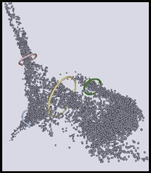
Intraoperative fluoroscopy during cardiac pacing lead implantation presents an occupational hazard to implanting physicians and their staff, both through cumulative radiation exposure and the orthopedic burden of wearing protective lead (Pb) garments. Pacemaker and defibrillator implantation is typically performed using fluoroscopy in a highly rationed manner due to the radiation hazard. We assessed preliminary feasibility of anatomic mapping and device lead implantation using an unrationed non-fluoroscopic 3D imaging system based on bioimpedance.
|
Stochastic Modeling and Simulation of Turbulent Reacting Flows, Sean Garrick and Victoria Interrante (2000) 9th International Symposium on Flow Visualization, Edinburgh.
[PDF]
[abstract]
|
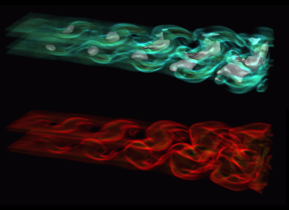
The filtered density function (FDF) is implemented for large eddy simulation of three-dimensional, planar jets under both non-reacting and chemically reacting conditions. In this methodology, the effects of the unresolved scalar fluctuations are taken into account by considering the joint probability density function of the scalar quantities in a stochastic manner. It is shown that under reacting flow conditions, the absence of closure for the SGS scalar correlations yields results which are significantly different from those obtained by the FDF. We visualize this data with a combination of volume and isosurface rendering, and introduce methods for reducing the memory and time costs that have historically precluded recording the results of the simulation at all calculated time steps.
|
Display Methods for Gray-Scale, Voxel-Based Data Sets, Victoria Interrante, William Oliver, Stephen Pizer and Henry Fuchs (1994) Three-Dimensional Confocal Microscopy: Volume Investigation of Biological Systems, John K. Stevens, Linda R. Mills and Judy E. Trogadis, eds., Academic Press, 1994, pp. 131-167.
[PDF]
[abstract]
|
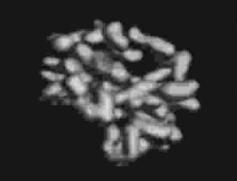
The dramatic increase in the use of 3D image acquisition devices over the past decade has inspired major new developments in the display of volume data sets. In this chapter we present an overview of these diverse display methods and discuss the relative advantages and disadvantages of each of the different approaches. In addition, we touch upon some of the major issues involved in creating high-quality images from volume data, including the problems of surface definition and object segmentation.
��������������������������������������������������������������������������������
|
Volume Rendering in Radiation Treatment Planning, Marc Levoy, Henry Fuchs, Stephen M. Pizer, Julian Rosenman, Edward L. Chaney, George Sherouse, Victoria Interrante and Jeffrey Kiel (1990) First Conference on Visualization in Biomedical Computing, pp. 4-10.
[PDF]
[abstract]
|
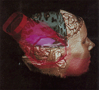
Successful treatment planning in radiation therapy depends in part on understanding the spatial relationship between patient anatomy and the distribution of radiation dose. We present several visualizations based on volume rendering that offer potential solutions to this problem. The visualizations employ region boundary surfaces to display anatomy, polygonal meshes to display treatment beams, and isovalue contour surfaces to display dose. To improve perception of spatial relationships, we use metallic shading, surface and solid texturing, synthetic fog, shadows, and other artistic devices. Also outlined is a method based on 3D mip maps for efficiently generating perspective volume renderings and beam掇-eye views. To evaluate the efficacy of these visualizations, we are building a radiotherapy planning system based on a Cray YMP and the Pixel-Planes 5 raster display engine. The system will allow interactive manipulation of beam geometry, dosimetry, shading, and viewing parameters, and will generate volume renderings of anatomy and dose in real time.
|
|
Three-Dimensional Display Methods for Image Data Obtained with SPECT, Benjamin M.W. Tsui, J. Lynne Hendricks, J. Randolph Perry, Victoria Interrante, Henry Fuchs and William H. McCartney (1989) European Journal of Nuclear Medicine, 15(8): 639.
[abstract]
|
We investigated various three-dimensional (3D) display methods for image data obtained with SPECT. The methods included cine display of reprojection images, 3D display of surface structures of the emission information, and simultaneous 3D display of emission and transmission information obtained with a SPECT system. Reprojection images are less noisy than the acquired projection data and can be obtained at any viewing angle. A 3D surface display of emission data is incapable of depicting deep-seated defects. The simultaneous display method is unique in providing simultaneous presentation of anatomical and functional information from the same patient without misregistration problems. Radioactivity distributions were obtained using standard SPECT imaging techniques (lung perfusion and T1-201 SPECT studies). Data for generating 3D anatomical surfaces were acquired using a transmission CT method on a SPECT system. The transmission study provides anatomical information not contained in the emission data. An emission image slice is superimposed on the corresponding transaxial cut-plane through the 3D anatomical surfaces. The resulting 3D model is displayed on a special graphics engine, which allowed interactive selection of the cut-plane and viewing angle. Choice of surface colors, transparency of skin surface, and shadowing is also possible. The simultaneous 3D display technique may be preferable for SPECT data because it better reveals the relationship of functional defects and anatomical landmarks.